In recent years, nanotechnology has been integrated into biomedical applications. Nanoparticles are efficacious in field of wound care, due their versatile therapeutic action in various stages of wound healing (Pérez et al, 2023). Their small size ranging between 1 to 100nm and large contact area increases the likelihood of bio-interaction and permeability at the wound surface (Gowda et al, 2023). Metallic based nanoparticles such as gold, silver, zinc and copper have been integrated in dressing products (Mendes et al, 2022). Copper nanoparticles (CuNPs) are widely used in dentistry (Ma et al, 2022), disinfectant industry (Alherek et al, 2023) and agriculture (Bakshi et al, 2021). Despite copper being a more accessible metal and more economic, the use of copper in wound care is relatively new with limited copper-based dressing available in the market.
Copper is essential trace element in living organisms, serving as a cofactor of enzymes necessary for cellular metabolism (Ruiz et al, 2021). Both copper insufficiency and excessive accumulation leads to a spectrum of diseases involving metabolic disorders, cardiovascular and neurodegenerative disorder (Balsano et al, 2018). In humans, the main source of copper is derived from intake of organ meat, nuts and shellfish. Copper is absorbed from the gastrointestinal tract and about 80% of it excreted through bile and faeces (Wapnir, 1998). The bioavailability of copper varies from 75% when consuming 400 mcg/day of copper through food to 12% when the diet contains 7.5 mg/day of copper (FNB: IOM, 2001).
The involvement of copper in the process of wound healing has been established through both in vivo and in vitro studies (Sandoval et al, 2022). During the inflammatory phase, the activity of neutrophils and macrophages are enhanced by Nerve Growth Factor (NGF). NGF promotes the growth of keratinocytes and the migration of fibroblasts during the proliferation and remodelling phase of wound healing (Sandoval, 2021). The activity of NGF is facilitated by the presence of copper ions (Kornblatt et al., 2016). Vascular Endothelial Growth Factor (VEGF) and angiogenin which are crucial modulators of angiogenic activity, are also regulated through copper-dependent stimulation (Sen et al, 2002).
The antimicrobial activity by copper is achieved through their adhesion to cell walls and cell membranes, leading to the release of copper ions. This in turn, disrupts enzymatic function, modifies protein structure (Xu et al, 2022) and gives rise to oxidative stress leading to cell death of susceptible microorganisms (Montero, 2019). CuNPs are shown to be effective in suppressing Pseudomonas aeruginosa biofilm formation (Lewis et al, 2015), as well as exhibit biocidal activity against multi-resistant organisms including MRSA, ESBL (Qiao et al, 2019) and Acinobacter baumannii (Al-Kadmy et al, 2023). Additionally, copper also facilitates generation of free radicals, which contribute to its virucidal effect (Merkl, 2021). Fungi, specifically Candida Albicans are found to be susceptible to copper ion fungicidal properties as well (Salah et al, 2021).
Prolonged exposure to high concentration of copper may cause cytotoxicity through the activation of reactive oxygen species (Karlsson et al. 2008). Zhou et al evaluated wound healing in vivo using different concentrations of CuNPs (10, 20, 50, 100 and 200µg/ml) and observe enhanced wound healing with no cytotoxicity. Considering the small size of CuNPs, any surplus particles are eliminated through the gastrointestinal system and efficiently metabolised within the body to ensure equilibrium is maintained. There is little evidence regarding the systemic effects of CuNPs through topical absorption, as most toxicology studies on CuNPs have focused on inhalation and ingestion route (ATSDR, 2022). However, copper has been used safely for decades as food additives, in intrauterine devices and dental amalgam. Copper based nanomaterials with maximum concentration of 1% are declared safe by Scientific Committee on Consumer Safety (2021) to be used on dermal and oral surface.
Given copper’s essential role in wound healing and its antimicrobial properties, it demonstrates great potential as a topical antiseptic agent. Regular cleansing with topical antiseptics has been an integral part of routine wound care. The integration of CuNPs in solution may aid the goal in wound bed preparation through its action as antimicrobial cleansing solution. Rinsing or soaking the wound will help in reducing microbial load and loosen devitalised tissue, facilitating debridement process (Rodeheaver and Ratliff, 2018). As slough, necrotic tissue, foreign bodies and excessive exudate can promote a favourable environment for wound infection and biofilm formation (Murphy et al, 2019). Ohsumi et al (1988) observed that the presence of copper ion (Cu2+) resulted in distruption of the plasma membrane’s permeability barrier in Saccharomyces cerevisae within a span of 2 minutes. There is also reduction of 99% of Escherichia coli and Staphylococcus aureus at 10 minutes of exposure to CuNP composites (Banerjee et al, 2023).
All wounds contain some degree of bacterial contamination, but this does not impact the course of wound healing (Edwards et al 2004, IWII 2022). However, when colonisation advances to wound infection, wound healing will be delayed. Davies et al (2007) associates wound with microbial load more than 1 X 10⁴ CFU/g to have this detrimental effect. Not all infection elicits symptoms or signs, thus, routine cleansing plays a crucial role in preventing and reducing microbial burden. Moreover, an expert panel concurred that proactive approach in preventive medicine need to be implemented in prevention of infection (Nair et al, 2023).
The objective of this study is to assess the clinical effectiveness of using nano-copper solution on wound bed preparation to enhance wound healing through re-epithelisation.
Methodology
This study was conducted in Wound Care Unit of Hospital Kuala Lumpur. Patients are selected randomly among the individuals who are regularly followed-up in the outpatient wound care clinic. The observation encompassed various size and types of wounds, including diabetic foot ulcers (n=4), surgical wounds (n=3), pressure injuries (n=2) and peripheral arterial ulcer (n=1). The study examines the wound healing progress over the period of 6 to 12 weeks using nano-copper solution containing 0.2% ionised copper (CuHeal++TM) as a wound cleanser.
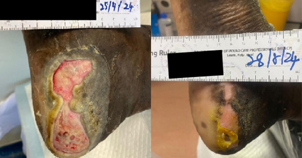
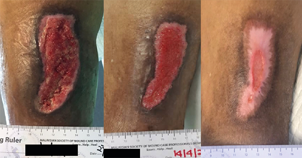
Each subject received the same standard of care and the principle of wound bed preparation were followed. The wound and peri-wound skin were cleansed using a solution containing nano-copper before applying gauze soaked with nano-copper solution on the wound for a period of 5 to 10 minutes. Then, debridement of wound is performed if indicated. Wound is cleansed again with nano-copper solution after debridement to remove any remnants of debrided tissue. Lastly, appropriate an wound dressing is applied based on wound assessment with TIME framework. The frequency of dressing changes differs depending on the condition of the wound and type of secondary dressing provided. However, all patients were subjected to the same procedure and documentation for subsequent follow-up. Data regarding the subjects’ demographic and their co-existing medical conditions are gathered along with the evaluation of wound progress. Consent had been obtained for all data and images collected during the period of study.
Discussion
Two subjects achieved complete wound epithelisation, whereas four subjects’ wound area achieved near complete closure. Patient no 7 and no 9 exhibit a lower percentage of wound size reduction in comparison to the others, possibly due to the substantial initial wound dimension (>100cm2) prior to commencement of treatment. However, all subjects (n=10) exhibited healthy, viable and moist wound bed at the end of study. The findings of the study align with multiple case reports on the use of copper-impregnated wound dressings to promote wound healing in challenging cases such as hard-to-heal wounds, diabetic foot ulcers, and infected wounds. (Melamed et al, 2023; Lu et al, 2022). Several studies have documented the role of CuNPs in promoting fibroblast proliferation and keratinocyte migration, both of which are crucial for wound repair (Wang et al, 2021, Kornblatt et al, 2016). Comparatively to a more widely used topical antimicrobial, silver, it has been established that dressing containing copper significantly promote angiogenesis and upregulates proangiogenic factors (Borkow et al, 2010).
During the period of study, none of the patient indicated signs of wound infection which supported the efficacy of nano-copper solution in reducing microbial load. Nair et al (2023) utilised fluorescence imaging to identify presence of bacterial on wound and they observed a decrease in bacterial load following wound cleansing with CuNPs based solution. Copper induces damage to bacterial cells mainly by the production of reactive oxygen species. On top of it, the inclusion of copper-containing nanoparticles induces a more potent antimicrobial action even at low concentration (Ma et al, 2022).
Not only effective as a wound cleanser, the nano-copper solution also proved useful to prevent wound infection and aid re-epithelisation. Ideally topical antiseptic should be able to reduce microbial load, promote wound healing, be non-toxic and not cause resistance (Bigliardi et al, 2017). The nano-copper solution applied topically, which is also odourless, colorless and sting-free is well tolerated by all subjects. No local or systemic side effects were reported despite the extended duration of usage up to 12 weeks in some subjects.
Undeniably, copper is indispensable in various human physiological function especially in context of wound healing. As an important mineral, the Recommended Dietary Allowance (RDA) for adults is 900µg/day. Copper deficiency is linked to reduction of collagen and elastin synthesis resulting in poor wound healing (Kornblatt, 2016). Additional research should be undertaken to explore the full potential of CuNPs in tissue repair and regeneration, other than as topical antimicrobial agent.
Limitations
This study has a limited sample size of 10, which is not representative of the general population. Furthermore, there was no specific delineation of patient selection criteria and the duration of study for each subjects varies depending on wound condition.
Conclusion
This case series concluded that the use of nano-copper solution in wound cleansing has synergistic effects that are of both antimicrobial and enhance wound healing without any adverse outcome.