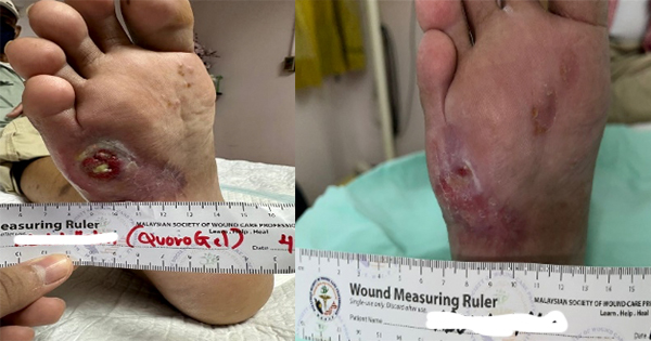
Category IV pressure ulcers (PU), the most serious stage of the classification system, are challenging to heal. The blood supply may have been cut off by thick layer of necrotic tissue and the wounds have often tunnelled through all the subcutaneous layer down to muscle and bone. The loss in tissue volume together with the amount of necrotic tissue delays healing and wound closure (Boyko et al, 2018; Headlam and Illsley, 2020).
While surgical debridement is usually necessary for optimal healing, the comorbidities of patients sometimes make this method unfeasible. Patients diagnosed with category IV PUs usually have poor mobility and/or are older people with associated long-term health conditions. It is not uncommon for the patient to be unfit or a have a high surgical risk. Poor mobility increases the difficulty in obtaining medical attention and lack of sensation in paralysed patients also limits the effectiveness of many conservative treatments. In these patient groups, an effective, dynamic on-site debridement treatment can significantly enhance the wound treatment process (Jaul, 2010).
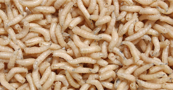
Maggot debridement therapy is well-recognised by healthcare professionals as an alternative treatment for hard-to-heal wounds (Mumcuoglu et al, 2001; Shi and Shofler, 2007; Gupta, 2008; Mohd Zubir et al, 2020). It is known as a reliable biological treatment for debridement in sloughy and necrotic tissues, acting by both mechanical and biochemical actions. Research suggests maggot debridement therapy can inhibit inflammatory responses, promote angiogenesis and fibroblast migration, it can also enhance the production of growth factors within the wound environment, all of which can help promote healing in hard-to-heal wounds (Prete 1997; Steenvoorde and Jukema, 2004; Horobin et al, 2005; van der Plas et al, 2007; Margolin and Gialanella, 2010; Sherman, 2014).
While surgical debridement allows faster removal of necrotic tissue from the patient, treatments such as maggot therapy offer a less traumatic solution to wound debridement. Maggot debridement therapy is also considerably faster and more dynamic than chemical debridement (Abdel-Samad, 2019; Mohd Zubir et al, 2020).
Our case report describes maggot debridement therapy conducted on an older patient with a category IV in their own home. The patient had poor mobility and the PU had been present for over a year.
Case report
The pateint was a 98-year-old Chinese male living in Hong Kong with a history of Parkinsonism, high blood pressure and was wheelchair bound. He was taking a long-term daily dose of Aspirin. The patient developed a PU over the thoraco-lumbar region more than one year ago due to general poor mobility and becoming bed bound after a chest infection. The wound was first recorded in April 2021, persisted for over a year, and was determined as a catergory IV PU with undermining of surrounding skin. The wound had been treated with chlostriopeptidase, which stimulates debridement and granulation, with minimal improvement. In April 2022 attempts were made to suture the wound together, which also failed.
The wound was first addressed clinically in October 2022 by sharp bedside debridement of necrotic tissue, followed by dressing changes, Aquacel (Convatec) and Cosmopor (Hartmann USA.inc), every 48 hours following the Community Nurse Service protocol. Cushioning and a ripple bed (an alternating mattress) for offloading were used. A barrier cream was applied to the wound. The patient was also turned regularly by nurses.
After a month, the treatment was taken over by a domestic helper, who followed the same protocol. No improvement was observed but a pocket had developed in the upper part of the wound. By the end of February 2023, (3 months later), the patient was hospitalised because of gastrointestinal haemorrhage. An air cushion bed provided by the hospital failed to prevent wound progression and the wound deepened after that. After the patient was discharged from the hospital, the wound was treated by private nurses. Attempts of surgical debridement had failed because of active bleeding. Chlostriopeptidase was employed for chemical debridement, which smoothed out some of the necrotic tissue, but the pockets of necrotic tissue remained unimproved (Figure 1).
Maggot debridement therapy of the wound began on 2 March 2023. At this time an open wound of around 12.7cm2 with necrotic tissue. There was extensive tunnelling under skin at the upper part of the wound. A deep pocket about 1.2cm was observed in the centre that had reached bone. Altogether five treatment cycles were performed, with each cycle lasting for three days. Before maggots introduction, Control gel formula dressing (CGF) dressing (Duoderm) was placed on the circumference of the wound. Medical maggots (Lucilia cuprina) were ordered from Cuprina, Singapore. In the first and second cycle the maggots were enclosed inside a permeable nylon bag. The biobags were placed directly onto the wound, followed by covering with three layers of dry gauze. The dressings were held in place with Omnifix (Hartmann USA.inc). At the end of the treatment cycle the dressings and the biobags containing maggots were discarded and the wound was washed with physiological saline. There was a three day gap between the first and second cycle because of logistic problem in importing maggots. During this gap chlostriopeptidase was applied and the wound was covered with dry gauze. (Figure 2)
Necrotic tissue on the surface of the wound had significantly reduced after the first two cycles. Tunnelling at the upper part of the wound had closed and the necrotic tissue in the flat area at the lower part of the wound had been cleaned out. Necrotic tissue was still present inside the pocket but had reduced in thickness (Figure 3). No negative response from the patient had been observed during this period.
The third cycle started on 10 March 2023. Targeting the pocket area, maggots were introduced directly onto the wound without containment. Maggots were washed out from the bag onto a gauze. The gauze containing the maggots was then directly applied to the wound that was then covered by three layers of dry gauze. After three days the maggots were washed out of the wound and discarded. New maggots were introduced following the same protocol after the wound was cleaned with physiological saline. After the third cycle there was another 24 hour gap caused by logistics issue. The wound was pulled together by Leukostrip (Smith and Nephew) and covered with dry gauze with no additional treatment during this period (Figure 4).
Continuous improvement in the condition of the wound was observed throughout the study. The maggots were completely removed having prevented the formation of necrotic tissue. At this point most of the wound area was replaced by fresh granulation tissue. The wound was showing signs of healing without using skin graft. During the 24 hours gap between the third and fourth cycle when the wound was pulled together with Leukostrip, the lower part of the pocket fused together and remain closed after removal of the pulling force. The remaining pocket also had a reduction in depth (Figure 5).
Mild swelling could be observed after the third cycle. The swelling disappeared during the gap period between cycles 3 and 4. The patient reported an itchy feeling after cycle 4. Skin redness was observed in the lower and left side of the wound outside the area covered by CGF dressing (Figure 6). These are likely caused by maceration from the wound moisture during the treatment. The redness disappeared within two days of treatment completion.
After cycle 5 the wound was cleared of necrotic tissue. Freshly grown granulation tissue was observed on the entire wound bed. The wound was covered by dermo-matrix (MatriDerm®, MedSkin Solutions Dr. Suwelack AG) for dermis regeneration (Figure 7).
Discussion
Mechanical debridement can be a challenging procedure in some patient groups. Older people with multiple comorbidities have high-anaesthetic/surgical risks. Patients on aspirin or other anti-platelet agents may also have significant bleeding. Deep wounds may develop to the bone meaning surgical debridement carries the risk of injury to spinal cord or nerve roots. Therefore there is a need for debridement methods with less surgical trauma. Among all the alternatives, maggot debridement therapy can achieve effective and dynamic debridement in the least traumatic way and within a reasonable timeframe.
As mentioned in the literature, maggot therapy can provide effective debridement, suppress inflammation and promote tissue regeneration in the same time (Sherman 2002; 2003; Gupta, 2008; Herwaldt and Rutala, 2015). The movement of maggots on the wound bed have a positive effect on the disruption of biofilm and stimulate surrounding tissue to promote faster regeneration. Furthermore, maggots can effectively absorb the wound discharge to maintain a clean environment for healing to take place. The merit of maggot debridement therapy, however, is based on the secretion of the maggots. Maggots secretion contain a mixture of proteolytic enzymes and chemicals including collagenase, allantoin, sulfhydryl radicals, protease, aminopeptidase, carboxypeptidases and growth stimulating substances (Abdel-Samad, 2019). These secretions not only digest and soften necrotic tissue on site but they also have antiviral and antibacterial effects that can disrupt the formation of biofilms. Together with the biochemical growth stimulation effects, maggot debridement therapy can help to establish a clean the wound bed for tissue regeneration without an invasive intervention. When compared with other non-invasive debridement methods, such as chemical debridement, maggot therapy provides a more dynamic and persistent debridement result. These make the therapy a potent substitute for surgical debridement.
In this case, the patient suffered from long-term health conditions that limit the choice of traditional treatments. Besides the patient’s old age and history of Parkinsonism, he is taking aspirin regularly and has bleeding tendency. Together with the location and condition of the wound, surgical debridement carried a high surgical risk. The patient had been treated for a long time with traditional conservative methods and advanced wound dressings with little improvement. As the wound progressed it reached the spinous processes making surgical debridement more difficult and riskier. Chemical debridement with chlostriopeptidase had shown positive effect in some areas of the wound but it failed to stop and reverse the continuous developing wound. Maggot debridement therapy on the other hand achieved a faster debridement, which is favourable for the healing process. It stopped the wound progression and led to recovery.
When compared with traditional surgical debridement, one major advantage of maggot therapy,observed in this case is that it can promote healing within the wound while debridement is ongoing. Beside removing necrotic tissue, maggots can also disgest and absorb fluid discharges from the surrounding tissue, preventing their accumulation. This provides a clean, solid and healthy wound bed for tissue regeneration. The healing progress is visually identifiable during the debridement process, with shrinkage in overall size and the disappearance of tunnelling. The pockets also significantly reduced in depth during the treatment.
One side effect is the skin redness, when maggots were directly applied to the wound. The redness was away from the wound, outside the area covered by CGF dressing. Judging from the location it is likely caused by maceration from the cumulation of moisture inside the gauze during the treatment. There is also mild redness and swelling at the circumference of the wound. This may be caused by allergy, but it cannot be confirmed without further study. All redness disappeared within two days after the termination of treatment.
Conclusion
Maggot debridement therapy achieved great success despite the delays in application due to logistical issues. There was steady improvement in the wound and the patient did not report any discomfort or show any major unwanted effects. Skin regeneration procedure could be conducted after 20 days of the therapy (5 cycle, 3 days each with 2 gaps). The case indicates the potential of maggot debridement therapy in treating category III/IV PUs in patients with underlying medical conditions, who are unsuitable for surgical operation. The therapy can improve the condition of the wound in a short period of time, which allows regeneration therapy to be conducted. Maggot debridement therapy can also be conveniently conducted outside hospital by a nurse, marking its potential in reaching a wider range of patients including older and people with mobility problem who have difficulty in travelling to hospital frequently.


