Hard-to heal wounds pose a significant challenge to both patient and healthcare providers as their healing process is often prolonged with conventional treatment (Beckmann et al, 2014). The usage of light therapy is not new and goes back to the ancient Egyptians and Indians who use sunlight (heliotherapy) for curative intent and attaining good health (Mosca et al, 2019).
Photo biomodulation (PBM) formerly known as Low level laser therapy (LLLT) is a painless treatment modality of chronic wound with minimal side effects (Mosca et al, 2019; Chen et al, 2022). Photo biomodulation refers to a form of light therapy using non-ionising forms of light sources such as lasers, light emitting diodes (LEDs) and broad-band light, in the visible and infrared spectrum, involving a non-thermal process with endogenous chromophores eliciting photophysical (i.e., linear and nonlinear) and photochemical events at various biological scales (Mosca et al, 2019). The latter involves a photochemical reaction that happens after photon absorption by a chromophore in the cell. Cytochrome c oxidase (CCO) is the main chromophore which takes in red and near-infrared (NIR) light while light-gated ion channels and channel rhodopsin are chromophores stimulated by blue and green light. One of the central wound healing pathways in photo biomodulation therapy is extracellular activation of transforming growth factor beta (TGF-β), which potentiates haemostasis (platelet derived TGF-β), inflammatory cells (macrophage-derived TGF-β and extracellular matrix (latent TGF-β-binding protein-associated TGF-β sequestered in the matrix; Mosca et al, 2019). Hence, faster re-epithelisation and reformed connective tissue, lower inflammatory process and increased proliferation of myofibroblasts were observed in wounds treated with LLLT (Mosca et al, 2019).
Objective
To assess effectiveness of photo biomodulation as adjunct to wound healing.
Methods
Patients attending wound care clinic Hospital Kuala Lumpur were randomly selected during routine day care wound dressing from June to December 2022 (Table 1). The wounds were of varying aetiologies.
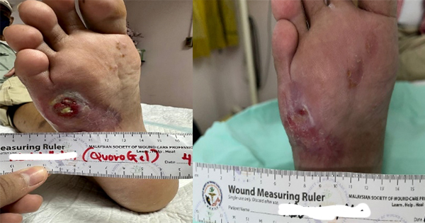
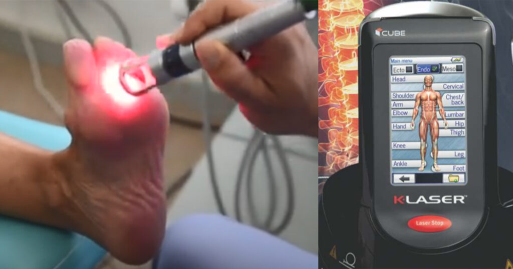
Before laser application, the wounds were cleaned and a primary dressing of either hydrogel, honey or silver alginate dressing were applied in accordance with the clinic’s usual standard of care. The wound dressings were changed at least twice a week while large wounds were dressed three times a week. Compression dressings were applied on several VLU patients. However, adherence was a major issue owing to discomfort. Offloading was applied to less than 50% patients as these patients had to apply for financial support to purchase offloading shoes. After wound dressing, laser therapy (K-CUBE 4 PLUS, VBS Medical Ltd) with four different wavelengths, 660, 800, 907 and 970nm at 30kJ, was applied via a handpiece held above the wound area for approximately three minutes The former was applied on each clinic visit (either every other day or twice a week) for three months. The wound size of each patient was measured at the beginning and end of the study.
Permission to use clinical images and case details for publication of this study were gained before the study.
Results
A total of 30 patients attending wound care clinic Hospital Kuala Lumpur were randomly selected during routine day care wound dressing from June to December 2022 (Table 1). These wounds were of varying aetiologies, 12 were diabetic foot ulcers (DFU), nine were venous leg ulcers (VLU) and nine were postoperative wounds.
The VLUs were present for at least six months before presentation at wound clinic, the DFUs duration ranged from three months to three years, while the postoperative wounds presented as early as two weeks to wound clinic.
Table 1 shows the percentage of wound size reduction after K-Laser treatment and Table 2 the percentage of wound reduction according to initial wound size.
Statistical analysis
Table 3 shows the initial wound size before the laser treatment has a mean value of 45.19cm2 and a standard deviation of 33.653cm2. Whereas, the final wound size after the laser treatment has a mean value of 20.94cm2 and a standard deviation of 25.541cm2.
Table 4 shows the paired samples correlations between initial wound size before laser treatment and final wound size after the laser treatment with the value of 0.733 at a p-value of 0.000.
Table 5shows the paired samples T-Test. In the tests, the null hypothesis is that the average of the differences between the paired observations in the two samples is zero. Conversely, the alternative hypothesis assumes that the true mean difference between the paired samples is not equal to zero. If the calculated p-value is less than 0.05, the conclusion is that, statistically, the mean difference between the paired observations is significantly different from 0.
Discussion
From this study, 66.7% patients (20 out of 30 patients) achieved at least 50% wound healing with six patients achieving 100% wound healing. The latter consisted of three DFUs, one VLUs and two postoperative wounds.
In terms of wound aetiology, DFUs accounted for half of the 20 patients achieving at least 50% wound reduction while only 8 postoperative wounds and three VLUs showed this similar outcome. Approximately half of the VLU patients achieved at least 30% reduction in wound size with K-Laser therapy. This is similar to the latest study findings by Rajhathy et al (2022), which reports 30% as basal healing rate for wounds managed with compression bandaging and advanced dressings.
Using the paired sample T-Test, the mean difference between paired observations (before and after laser treatment programme) is significantly different from 0. The finding from the results indicates that there is a mean difference between the two groups and the p-value is less than 0.05. Hence, the wound size of patients has reduced after the laser treatment.
The device used in this case series is K-Laser with Class IV laser system which emits light with wavelengths of 660, 800, 905 and 970nm. The energy delivered by this device can be tailored according to chronicity, skin shade, and depth of penetration (Nair et al, 2021).
Laser therapy accentuates wound repair in the preliminary stages of wound healing i.e. faster proliferation phase indicated by intensive fibrillogenesis and angiogenesis, as well as better regulation of intricacies of wound healing by cytokine regulation (Pavlovet al, 2020). Other beneficial effects associated with photo biomodulation include enhancing tissue renewal, controlling inflammation, reducing pain and oxidative stress. This can be explained by its ability to stimulate mitochondria, increasing adenosine triphosphate (ATP) production and the downstream release of growth factors. These growth factor bind to cell surface receptors and induce transcription of genes for increased cellular proliferation, viability, and migration in numerous cell types, including stem cells and fibroblast (Nair et al, 2021). K-Laser therapy significantly increases the formation of new capillaries, therefore increases tissue oxygenation and accelerating healing processes (Nair et al, 2021).
Limitations
One limitations of this study was that a certain degree of selection bias was present as most patients had undergone surgical debridement before wound care visit. Furthermore, venous leg ulcer patients were mostly non-concordant with wearing compression bandaging due to the discomfort, which will affect the healing rates.
Conclusion
This case series has demonstrated that usage of photo biomodulation as adjunct to wound healing resulted in significant reduction in wound size (p<0.05). This can be explained from its effects in enhancing proliferation, tissue renewal, controlling inflammation and oxidative stress. Hence, more high-levelled trials i.e randomised study should be conducted to incorporate K-Laser in clinical protocols of wound management (Mosca et al, 2019).