A non-healing wound is defined as a wound that has not healed by 40–50% after four weeks of good standard of care (Atkin et al, 2019). It is often associated with patients with underlying medical conditions such as diabetes and vascular diseases, as well as older people (aged 60 and above; Hall et al, 2015). The majority of patients with non-healing wounds frequently report pain as the most burdensome symptom (Mustoe, 2004; Iqbal et al, 2017). This pain often results in reduced productivity and quality of life (QoL), in addition to the significant economic impact of managing these wounds (Guest et al, 2015; Sen, 2021). Hence, there is an urgent need to implement effective novel treatments to promote faster wound healing. It has been identified that photobiomodulation therapy (PBMT) has shown promising results (Cicchi et al, 2016).
The North American Association for Photobiomodulation Therapy (2021) coined the term ‘photobiomodulation’ in 2014, defining it as a form of light treatment eliciting photophysical and photochemical events at various biological scales. Blue light (410–430nm) within the visible light spectrum can promote wound healing in both acute and chronic wounds. This occurs through photophysical and photochemical effects, ultimately leading to the application of PBMT.
Photochemical effects play a more crucial role in the healing of non-healing wounds because these wounds often suffer from poor blood flow. Blue light’s action on cytochrome C/cytochrome C oxidase, activation of flavins and the increase in reactive oxygen species become particularly significant (Magni et al, 2020; Rossi et al, 2021). These reactive oxygen species, in turn, can induce controlled inflammation, a process highlighted in the Blue Light for Ulcer Reduction study. The study demonstrated that treating non-healing wounds of varying aetiologies with blue light for 60 seconds once a week in addition to standard care significantly enhances re-epithelialisation (Fraccalvieri et al, 2022). Notably, there was a statistically significant difference between PBMT and standard care alone, with this effect appearing particularly pronounced in venous leg ulcers (VLUs; Fraccalvieri et al, 2022). Blue light also has the ability to regulate the activity of fibroblasts, which are responsible for producing extracellular matrix and collagen during tissue remodelling (Cicchi et al, 2016).
PBMT therapy can facilitate tissue repair by increasing capillary and vascular circulation, stimulating cellular respiration and increasing fibroblast activity and collagen deposition (Priola et al, 2021). Specifically, it has been found that blue light (410–430nm) therapy (EmoLED) is effective in promoting wound healing and reducing pain in patients with chronic VLUs, cutaneous vasculitis, and traumatic ulcers that were not responding to standard treatments (Mosti and Gasperini, 2018; Marchelli et al, 2019; Dini et al, 2021).
In this case series, we present observations derived from the treatment of EmoLED in nine patients diagnosed with non-healing wounds of the lower limbs of different aetiologies, such as diabetic foot ulcers (DFUs), VLUs and traumatic wounds.
Material and methods
The study included nine consecutive patients diagnosed with non-healing wounds of various aetiologies in the lower limbs that were not responding to standard care for an average of 57 days before therapy. Patients were enrolled from the Specialist Clinic Ambulatory Care Centre, Hospital Kuala Lumpur, Malaysia (SCACC HKL). Exclusion criteria included patients with neoplastic lesions or those known or diagnosed with porphyria.
After conducting standard wound hygiene procedures, including thorough cleansing of the wound with distilled water and, if necessary, debridement and reshaping, all patients received EmoLED treatment. For patients with VLUs and DFUs, a 120-second weekly treatment was administered to the affected area, while other wound types received a 60-second treatment per wound area. Following each treatment, wounds were managed according to standard care protocols using modern dressing techniques. DFU cases involved offloading, while VLU patients received compression therapy.
The LED irradiation system used is a portable device emitting blue light (400–430nm), provided by EmoLED Srl (Florence, Italy). The EmoLED blue LED light device illuminates a circular 5cm diameter area at a distance of 4cm from the wound bed for either 60 or 120 seconds. This results in a power density of 120mW/cm2 LED radiation, corresponding to an energy density dose of 7.2J/cm2 (60 seconds) or 14.4 J/cm2 (120 seconds).
Ethical considerations
All patients that were enrolled in this study provided written consent for both the therapy they received and the use of their health record data, including their medical history, blood investigations and photographs.
Results
This study reports the outcome of nine patients who were treated with EmoLED and successfully achieved complete wound healing. Table 1 provides a summary of each patient’s comorbidities and length of time taken to achieve complete wound healing.
The patient’s ages ranged from 45 to 83 years. Of the nine patients, seven had diabetes mellitus, six of which also had vascular diseases.
On average, patients had their wound for 62 days before commencing EmoLED therapy.
However, this is skewed by case 5, as the duration of wound was calculated post surgery as it is a different wound from his DFU wound, which means the average duration is higher than reported. The average time taken to achieve complete wound healing is 109 days from the beginning of EmoLED treatment.
Case 1
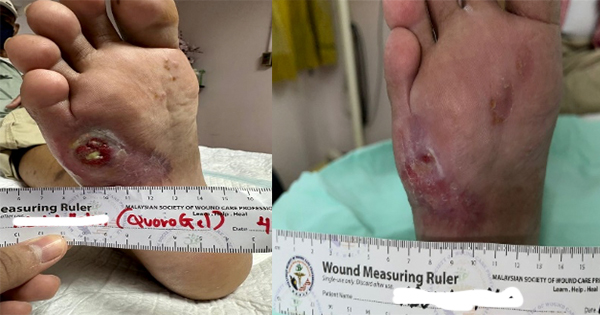
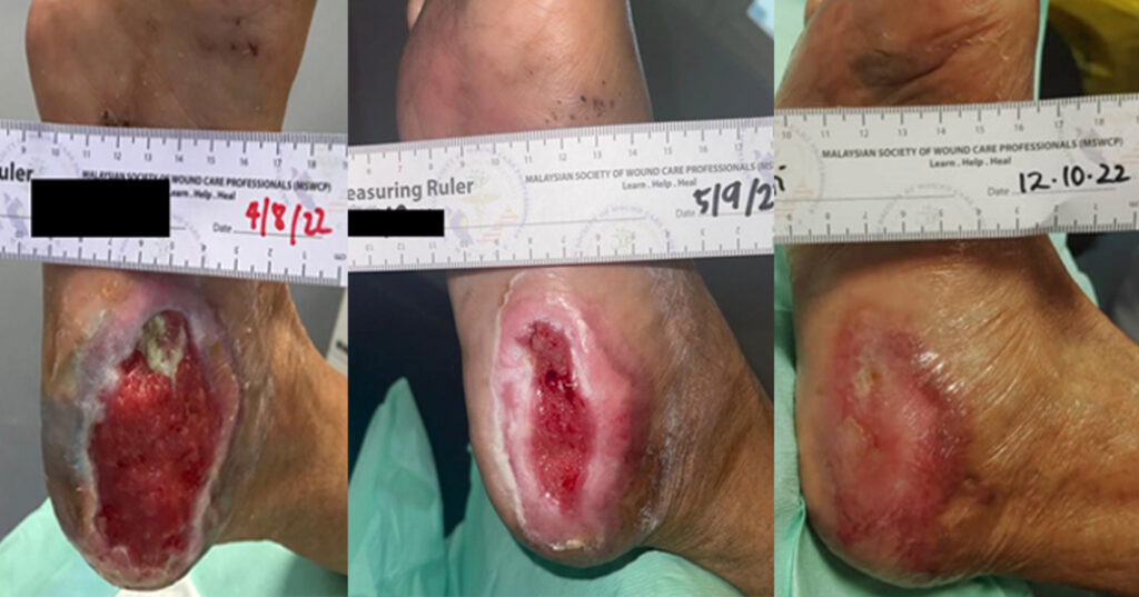
A 58-year-old Malay female with underlying poorly controlled diabetes mellitus developed a DFU on her right heel in May 2022 (Case 1). The patient began standard wound treatment at SCACC HKL on 7th June 2022; however, she noticed that wound healing was delayed. The wound assessment revealed a 9cm x 5cm wound on the plantar aspect of the right heel, extending to the medial edge of the foot. The wound bed was noted to contain 15% slough and 85% granulation tissue, with a moderate to high level of exudate. The caudal part of the wound edge was slightly macerated, while the other segments appeared healthy. The periwound skin was normal. On 4 August 2022, the patient was enrolled in EmoLED treatment, receiving one session per week lasting 120 seconds. Complete wound closure was achieved on 12 October 2022.
Case 2
A 47-year-old Malay female, with underlying, poorly controlled diabetes mellitus, developed a DFU on her left foot in May 2022 (Case 2). During this month, she underwent Ray’s amputation of the left 2nd and 3rd toes and extensive wound debridement of the left foot. Her standard wound treatment at SCACC HKL began on 9 June 2022, involving modern dressing and wound hygiene protocols. However, there was no significant improvement observed.
She was subsequently enrolled in EmoLED treatment starting on 8 August 2022. The wound assessment revealed a 30cm x 9cm wound with 20% slough, 70% granulation tissue and 10% epithelialisation. Exudate levels were moderate and the wound edges and periwound skin appeared healthy. The patient received EmoLED irradiation once a week, with each session lasting 120 seconds.
Complete wound closure was achieved on 20 December 2022.
Case 3
A 70-year-old female patient of Indian ethnicity, with underlying diabetes mellitus, hypertension, dyslipidaemia and ischemic heart disease, underwent coronary artery bypass graft surgery on 17 May 2022. The right long saphenous vein was harvested for the procedure; however, the wound on her right leg healed poorly (Case 3). She was referred to SCACC HKL and commenced EmoLED treatment on 26 July 2022.
The wound assessment revealed three separate wounds, each with different dimensions: 4cm x 2cm, 7cm x 3cm, and 7cm x 2cm, with 10% slough, 80% granulation tissue and 10% epithelialisation. A moderate amount of serous exudate was present and wound edges and periwound skin appeared healthy and normal. A standard dressing regimen was applied to the area, using distilled water for cleansing during each visit. The wound area received EmoLED irradiation once a week, with 60-second sessions.
Complete wound healing was achieved on 27 September 2022.
Case 4
An 83-year-old male of Indian ethnicity with underlying diabetes mellitus, hypertension and ischaemic heart disease, presented with complaints of pain and swelling in his left foot. The wound on his foot had pus and emitted a foul smell. He underwent incision and drainage and was diagnosed with a DFU in May 2022 (Case 4). An X-ray was performed on the left foot, but it did not reveal any evidence of osteomyelitis changes. Oral antibiotic treatment was started, along with a regular dressing regimen, but no significant improvement was shown. He was subsequently referred to SCACC HKL and started the EmoLED treatment on 29 July 2022.
Wound assessment showed a 2cm x 2cm wound with 80% slough and 20% granulation tissue. The wound had a moderate amount of serous exudate and minimal undermining edge. The periwound skin was normal. A standard dressing regime was applied to the area, cleansing it with distilled water during each visit.
The wound was irradiated with EmoLED once a week, with sessions lasting 120 seconds.
Complete wound healing was achieved on 02 December 2022.
Case 5
A 51-year-old man of Indian descent, with underlying diabetes mellitus, hypertension, and dyslipidaemia, developed a right DFU in 2021. He had been receiving daily dressing changes at a primary care clinic. However, on 22nd July 2022, he underwent above-the-knee amputation due to an infected right DFU with necrotising fasciitis (Case 5). His angiography revealed a critical narrowing up to the popliteal bifurcation Because of his long history of poor wound healing, the patient was referred to SCACC HKL and started EmoLED treatment on 2nd August 2022.
Upon assessment, the wound measured 12cm x 15cm and had 20% slough and 80% granulation tissue. There was a moderate amount of exudate and the wound had minimal undermining at the edges. The periwound skin appeared normal. Standard dressing regimen was applied to the area, with cleansing using distilled water during each visit.
The wound was irradiated with EmoLED once a week with 120-second sessions.
Complete wound healing was achieved on 3 November 2022.
Case 6
A 45-year-old female of Malay ethnicity with underlying diabetes mellitus, hypertension and stage 3 chronic kidney disease was diagnosed with an infected right DFU in April 2022. The patient underwent Ray’s amputation of the right big toe on 3 May 2022 and was referred to the wound care clinic.
Foot assessment showed a normal ankle brachial pressure index (ABPI) of 1.0 without signs of neuropathy. The wound measured 8cm x 6cm with 20% slough and 80% granulation tissue (Case 6). Exudate levels were moderate, and the lateral edge of the wound was macerated. Periwound skin was normal. A standard dressing regimen was applied to the area, with cleansing using distilled water during each visit. However, there was no significant improvement with the dressing used. Therefore, starting from 3rd August 2022, the wound was irradiated with EmoLED once a week, with each session lasting 120 seconds.
Complete wound healing was achieved on 07 December 2022.
Case 7
An 82-year-old female patient of Chinese ethnicity with underlying hypertension and osteoarthritis, developed a VLU on her left lower limb in July 2022 (Case 7). The patient was referred to SCACC HKL and began the EmoLED treatment on 5 August 2022.
The wound assessment revealed two ulcers, one measuring 2cm x 2cm and the other 2.5cm x 2cm, located on the medial aspect of the shin near the ankle. The wound bed consisted of 80% slough and 20% granulating tissue. There was a moderate amount of exudate with a healthy wound edge, and the periwound skin appeared normal. A standard dressing regimen was applied to the area, with cleansing using distilled water during each visit. The wound received EmoLED irradiation once a week, with sessions lasting 60 seconds.
Complete wound healing was achieved on 03 October 22.
Case 8
A 70-year-old male of Chinese ethnicity, without any underlying comorbidities, sustained an injury with a sharp metal object on his right shin on 27 July 2022 (Case 8). Haemostasis was achieved at the emergency department, and subsequently, the patient was referred to SCACC HKL and began EmoLED treatment on 15 August 2022.
Upon assessment, the wound measured 9cm x 5cm and had 90% slough with 10% granulation tissue. There was a low amount of serous exudate, and the wound had a healthy edge. The periwound skin appeared normal. A standard dressing regimen was applied to the area, with cleansing using distilled water during each visit.
The wound was irradiated with EmoLED once a week with 60-second sessions. Complete wound healing was achieved on 16 November 2022.
Case 9
A 64-year-old male patient of Malay ethnicity, with underlying diabetes mellitus and ischemic heart disease, was diagnosed with right DFU in March 2022. Due to wound infection (Case 9), he underwent wound debridement on 1 July 2022 and initiated EmoLED treatment at SCACC on 14th July 2022.
Upon assessment, the wound measured 15cm x 8cm. Using the TIMES concept for wound assessment, 80% of the wound bed was covered in slough and 20% granulation tissue. There was a slight malodour, and the wound had a moderate amount of serous discharge. The wound edges appeared healthy, and the periwound skin was normal. After cleansing the wound, EmoLED was applied for 120 seconds, followed by a standard dressing procedure with hydrogel as the primary dressing. Dressing changes occurred every other day, while EmoLED was applied once a week during this period.The wound completely healed and the patient was discharged on 7th December 2022.
Discussion
SCACC HKL serves as the referral centre for wound management, and all the patients involved in the study received wound care that adhered to the best standard practices before the initiation of PBMT. For patients with DFUs, a schedule of wound dressing sessions every other day was followed, involving the following steps:
1. Wound cleansing: The first step involved cleansing the wound using distilled water
2. Debridement: When necessary, debridement was performed to remove unhealthy tissues and biofilm. This could be achieved through mechanical methods using gauze or a curette, or through sharp debridement with a blade. Refashioning of the wound edges was also carried out if they were macerated or callused
3. Primary dressing: Primary dressing options were selected based on the specific wound condition. Hydrogel was used to maintain the balance of wound moisture, antimicrobial dressings such as silver were employed in case of infection, and foam or hydrofiber dressings were used if there was a significant amount of exudate. These methods were adjusted based on the wound assessment conducted during each session
4. Offloading: Offloading was recommended, especially for wounds on the plantar aspect. This was achieved through adequate padding or casting, while non-weight-bearing methods were used for other cases.
For patients with VLUs, a compression bandage system was applied. However, despite being classified as non-healing wounds, these patients did not progress according to the expected timeline of normal wound healing
In this pilot study for our Blue Light Empirical Shine study, it has been shown that despite having different underlying causes, all nine patients responded positively to blue light therapy. This suggests that all wounds may share a common pathophysiological pathway, which results in similar responses to blue light and ultimately leads to the same outcome—complete wound healing.
Limitation
To improve the validity of the result, a double-blinded randomised controlled trial should be conducted.
Conclusion
Our preliminary observations indicate that light therapy holds promise as a novel treatment approach for expediting the healing of non-healing wounds. It has demonstrated the potential to reduce wound size, alleviate pain and improve the rate of wound healing. To substantiate these findings and establish the optimal treatment dose and duration, we intend to undertake a more extensive study.
We believe that blue light therapy could have a significant impact on the QoL of patients with chronic wounds and indirectly contribute to the sustainability of the healthcare economy.