Chronic wounds represent a significant burden to healthcare systems, particularly among patients with diabetes and chronic venous disease (Guest et al, 2020). Delayed wound healing is often multifactorial, involving impaired perfusion, infection, neuropathy, repeated trauma, and poor tolerance of or adherence to standard therapies, such as debridement, offloading, or compression (Armstrong et al, 2017; Harding et al, 2015).
DermaRep® (Biovotec) is a sterile, single-use, dissolvable wound dressing supplied as a thin, flexible sheet composed of purified eggshell membrane protein. The sheet has a soft, translucent, collagen-like appearance and can be easily trimmed to match the size and shape of the wound bed. It contains collagen, glycosaminoglycans and other bioactive components that may support cellular migration and tissue regeneration.
Purified eggshell membrane protein contains naturally occurring collagen, glycosaminoglycans, elastin, hyaluronic acid and other extracellular matrix components that are involved in tissue repair. These components may support cellular attachment, migration, angiogenesis and granulation tissue formation while maintaining a moist wound-healing environment. As a bioresorbable scaffold, DermaRep®® is intended to integrate gradually into the wound bed, providing structural support for tissue regeneration without requiring traumatic removal during dressing changes (Hoghooghi et al, 2020; Singer and Clark, 1999).
The product is designed for straightforward clinical application. Following standard wound bed preparation (including cleansing and, where appropriate, debridement), the DermaRep® sheet is applied directly to the wound surface. The material conforms closely to the wound bed and hydrates with wound exudate, gradually integrating into the tissue as it dissolves. A secondary dressing is required to secure the product in place and manage exudate, selected according to wound characteristics. The product is intended to remain in situ and does not require removal of residual material at dressing changes because it is bioresorbable. It may be layered or reapplied as clinically indicated. The frequency of application depends on wound exudate levels and clinician preference; however, DermaRep® is designed to remain in place between dressing changes and does not require routine removal.
This case series presents real-world clinical experience using DermaRep® in a range of chronic wound aetiologies, highlighting its ease of use and potential role as an adjunctive therapy in complex, non-healing wounds.
Methods
This is a descriptive case series conducted at the Wound Care Unit, Hospital Kuala Lumpur. Adult patients with chronic non-healing wounds were included based on clinical indication for advanced wound dressing use. DermaRep® was applied as part of standard wound care. Dressing frequency and adjunctive treatments, including offloading, compression therapy and specialist referrals, were individualised according to wound type and patient tolerance.
Wound dimensions (length × width) were recorded at baseline and subsequent follow-up visits. Outcomes assessed included wound size reduction, time to healing, and clinical progression of granulation and epithelialisation.
Ethics and consent
Ethical approval was not required for this descriptive case series. Written informed consent was obtained from all patients for the use of anonymised clinical data and wound photographs for educational and publication purposes.
Case series
Case 1: Diabetic foot ulcer
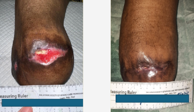
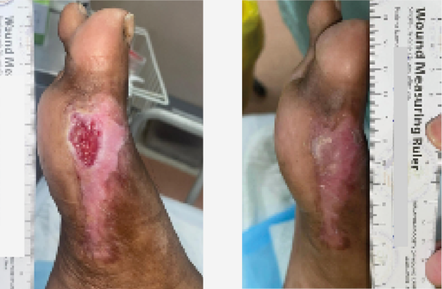
A 64-year-old Malay man with diabetes and hypertension presented with a diabetic foot ulcer (DFU) following ray amputation of the right fifth toe, which had remained static for 2 months. The wound measured 3 cm × 2 cm at baseline. Following DermaRep® application, progressive wound reduction was observed, with complete epithelialisation achieved by day 18 [Figure 1].
Case 2: Diabetic foot ulcer
A 47-year-old Malay man with poorly controlled diabetes presented with a chronic right DFU of approximately 3 months’ duration following surgical debridement. Minimal wound progress was observed despite 4 months of standard wound care at the Wound Care Unit, Hospital Kuala Lumpur, including regular dressing changes and offloading. Baseline wound size was 5 cm × 4 cm. The wound demonstrated steady improvement and achieved complete healing by week 9 [Figure 2].
Case 3: Venous leg ulcer
A 62-year-old Indian woman with obesity and varicose veins presented with a painful non-healing left lateral shin ulcer of 6 months’ duration. The patient was unable to tolerate debridement. Despite this, with the use of DermaRep®, the wound size reduced from 4 cm × 3 cm to complete closure by week 10 [Figure 3].
Case 4: Recurrent diabetic foot ulcer
A 42-year-old Malay woman with diabetes, hypertension and dyslipidaemia developed a recurrent left DFU following ray amputation. Baseline wound size was 3 cm × 3 cm. DermaRep® was initiated, and the wound healed completely by day 25 [Figure 4]. Her adherence to offloading was improved.
Following wound care education and prescription of specialised diabetic footwear, the patient demonstrated improved adherence to offloading recommendations.
Case 5: Large venous leg ulcer
A 63-year-old Malay man with diabetes and dyslipidaemia presented with a large left lower leg venous ulcer measuring 18 cm × 8 cm. Previous negative-pressure wound therapy and split-thickness skin grafting had failed. DermaRep® was introduced, and the wound was reduced to 8 cm × 4 cm by week 11 [Figure 5]. At the end of the observation period, the wound continued to demonstrate progressive healing; however, complete closure had not yet been achieved.
Case 6: Recurrent venous leg ulcer
A 69-year-old Malay man with multiple comorbidities presented with a recurrent right lower limb venous ulcer. Baseline wound size was 9.6 cm × 6 cm. Using DermaRep® and with good adherence to compression therapy, the wound reduced to 6 cm × 4 cm within 4 weeks [Figure 6]. Following the observation period, the patient was referred to the Vascular Surgery team for further vascular assessment and to the Dermatology team for skin assessment. The patient demonstrated good adherence to compression therapy, which was continued as part of the ongoing management plan.
Case 7: Diabetic foot ulcer
A 59-year-old Malay woman with diabetes and dyslipidaemia presented with a large left DFU following ray amputation. Over 11 weeks of DermaRep® therapy, the wound demonstrated progressive size reduction and improved granulation tissue formation [Figure 7].
At the end of the observation period, the wound demonstrated ongoing granulation tissue formation and progressive size reduction. The patient remained under follow-up and had been referred to the Plastic Surgery team for consideration of split-thickness skin grafting (SSG).
Case 8: Diabetic foot ulcer
A 77-year-old Malay woman with diabetes and hypertension presented with a right diabetic foot ulcer following surgical debridement. Wound dimensions reduced steadily over an 18-day period of DermaRep® application [Figure 8]. Following the 18-day observation period, the patient was unable to continue follow-up at our centre because of travel distance and was referred to a healthcare facility closer to home for ongoing wound management.
Results
Five patients had DFU, and three had venous leg ulcers. Five wounds achieved complete healing, and the remaining three demonstrated substantial wound size reduction and healthy granulation tissue formation following DermaRep® application [Table 1].
Complete wound closure was achieved in five cases within 18 days to 11 weeks. The remaining cases showed size reduction with healthy granulation and epithelialisation. No dressing-related adverse events were observed.
Discussion
This case series highlights the potential role of DermaRep® as an adjunctive dressing in complex chronic wounds. Notably, favourable outcomes were observed in patients unable to tolerate debridement and in wounds with prior treatment failure. The dissolvable nature of the dressing may contribute to reduced pain and improved patient acceptance (Singer and Clark, 1999).
Limitations include the small sample size, heterogeneity of wound aetiologies, and lack of a comparator group. Follow-up duration varied between cases because the observation period ended before all wounds had achieved complete closure. In addition, one patient was unable to continue follow-up at our centre and was referred to a clinic closer to home for ongoing wound care. Nevertheless, the findings provide preliminary evidence supporting further evaluation in controlled studies.
While causality cannot be established due to the descriptive nature of this series, the results are consistent with emerging evidence supporting the role of advanced biologically active dressings in chronic wound care. Further prospective studies, including comparative and randomised designs, are required to better define the clinical effectiveness and cost implications of DermaRep®.
Limitations
The main limitations of this case series include its descriptive design, small number of patients, absence of standardised outcome measures, and limited generalisability. These limitations are common to early clinical evaluations and observational case series in wound care research (Zelen et al, 2013).
Conclusion
DermaRep® dissolvable wound dressing demonstrated encouraging clinical outcomes in a range of chronic and complex wounds, including diabetic foot ulcers and venous leg ulcers. The dressing was well tolerated and associated with progressive wound healing, even in challenging clinical scenarios. Larger prospective studies are warranted to further evaluate its effectiveness and role within standard wound care pathways.
Acknowledgements
The authors thank the Wound Care Team, Hospital Kuala Lumpur, for their support in patient management and data collection. Products were provided by DermaRep®.