Wound healing depends not only on the condition of the wound bed, but also the integrity of adjacent periwound skin. A consensus document by LeBlanc et al (2021) defined periwound skin as being the area around a wound that may be influenced by variable factors and underlying wound pathology. Periwound skin plays an essential role in maintaining skin barrier function, modulating inflammation and facilitating epithelial migration during wound repair (Ho and Kupper, 2019; Rousselle et al, 2019). A disruption to this zone, due to maceration, desiccation, hyperkeratosis, contact dermatitis or excoriation, could compromise the cutaneous barrier and predispose the wound to risk of infection, delayed healing and pain (Woo et al, 2017; Holloway and Mahoney, 2021).
In vitro studies using organotypic skin cultures and scratch assays have demonstrated that epithelialisation originates from the intact surrounding epidermis rather than the wound bed itself (Safferling et al, 2013; Castellano-Pellicena and Thornton, 2020). Exposure of keratinocytes to chronic inflammatory cytokines, proteases, or barrier disruption impairs cell migration and differentiation, resulting in cellular senescence observed at chronic wound edges (Pastar et al, 2014). These findings highlight the importance of healthy periwound skin for a successful wound closure.
In vivo evidence from animal and human studies further supports the key role of maintaining a good skin barrier. Murine and porcine wound models show that excessive inflammation or barrier disruption in perilesional skin delays epithelialisation and causes excessive scarring (Eming et al, 2007).
Compromised skin integrity also leads to moisture loss due to increased transepidermal water loss (TEWL). TEWL is recognised as a dependable indicator of epidermal barrier integrity (Jansen van Rensburg et al, 2019). Increased TEWL indicates a functional disruption of the epidermal barrier, which is linked to inflammation and a higher incidence of wound recurrence (Chattopadhyay et al, 2024).
Using a murine model, Roy et al (2014) revealed that compromised epidermal barrier function, increased transepidermal water loss and inflammation impair wound healing and delay functional closure, despite apparent surface epithelialisation. However, failure to restore the skin barrier integrity renders the healed area susceptible to recurrence, highlighting the necessity of functional wound closure beyond the visual wound closure (Sen and Roy, 2021).
Despite the increasing recognition of integrating periwound management in wound healing, most clinical interventions focus primarily on wound bed preparation, with limited studies examining interventions for protection of the periwound microenvironment.
Mesenchymal stem cell–conditioned media (MSC-CM), the acellular secretome of mesenchymal stem cell, has garnered growing interest for its therapeutic benefits (Trigo et al, 2025). MSC-CM contains abundant components including cytokines, interleukins, growth factors, exosomes, lipid mediators and cell adhesion molecules, which are involved in the paracrine signalling pathway as part of tissue repair (L et al, 2019). Its utilisation in regenerative medicine has been applied across diverse tissues and organs, such as skin, cartilage, bone, lungs, liver, brain and heart (Bogatcheva and Coleman, 2019). Riedl et al (2021) have provided a comprehensive overview of mesenchymal stromal cells and their secretome in enhancing keratinocyte migration, modulates inflammation, and supports extracellular matrix remodelling, all of which are critical for wound repair.
Therefore, the targeted application of MSC-CM based therapy to periwound skin is a viable alternative to restore epidermal barrier and facilitate wound healing.
Methodology
This study was conducted as an observational case series describing clinical outcomes following the use of MSC-CM skin lotion (Sollagen, Thermo Fisher Scientific) on periwound skin. The MSC-CM is derived from red deer umbilical cord lining.
All patients provided informed consent for treatment and use of data for reporting purposes. Demographic data, wound characteristics, and clinical outcomes were extracted from medical records and wound documentation. The study adhered to the principles of the Declaration of Helsinki and was approved by hospital’s review board.
A total of 20 patients with various wound aetiologies were included. Patients were recruited via convenience sampling from a wound care clinic. Inclusion criteria were:
- Adults aged ≥18 years.
- Presence of a wound with intact periwound suitable for topical application.
- Able to attend every 2 days for regular follow-up appointments.
Exclusion criteria were:
- Malignancy at the wound site.
- Clinical signs of untreated or spreading infection.
- Fungal skin infection
- Skin maceration.
- Known allergy to product components.
Topical lotion containing MSC-CM was applied exclusively to the periwound skin within 4 cm of the wound edge and not in contact with the wound bed. Application frequency was standardised to once daily. All patients received standard wound care, including wound cleansing, debridement where indicated and adjunctive therapies such as compression or offloading as clinically required.
The primary outcome was percentage reduction in wound area over time, calculated using serial wound measurements.
Wound assessment and wound bed preparation was performed at each visit with the TIME (T-Tissue Management, I-Inflammation and infection control, M-Moisture balance, E- Epithelial edge advancement) framework (European Wound Management Association, 2004). Secondary outcomes included changes in periwound skin using a standardised tool, the Harikrishna Periwound Skin Classification system [Table 1] (Nair et al, 2020), time to wound closure where applicable and occurrence of adverse events related to topical application. The study duration was 8 weeks.
Results
Twenty patients with chronic wounds were included in this observational case series. The median age was 56.5 years, with a predominance of male patients (75%). All patients had diabetes, reflecting a high-risk wound population. Wound aetiology was evenly distributed between diabetic foot ulcers (n=10, 50%) and other wounds (n=10, 50%) such as surgical site infections (n=3), carbuncles (n=3), venous leg ulceration (n=3) and burns (n=1). The median wound duration at baseline was 8.0 weeks.
At baseline, the median wound area was 27.0cm². At endpoint assessment, the median wound area reduced to 4.0cm². This corresponded to a median percentage wound area reduction of 84.7% [Table 2].
Seven wounds (five diabetic foot ulcers and two surgical site infections) achieved complete closure by the endpoint of 8 weeks, while others demonstrated substantial reduction in size. No cases exhibited an increase in wound area during the study period. Individual wound size reduction varied, but a consistent overall trend towards wound closure was observed [Figure 1].
Baseline periwound skin assessment using HPSC highlighted that, in most wounds (n=19) with periwound damage, this was a result of desiccation or inflammation [Figure 1]. An overall reduction in median wound area was observed from baseline to endpoint, indicating overall improvement [Figure 2]. At the endpoint, marked improvement in the periwound skin condition was observed, with 18 cases (90%) classified as Class 0 (healthy) and the remaining 2 cases (10%) as Class 1 (at risk), as seen in Figure 3. No cases demonstrated worsening of periwound skin status, and no adverse events related to periwound application of MSC-CM lotion were reported during the treatment period.
Case studies
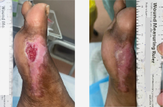
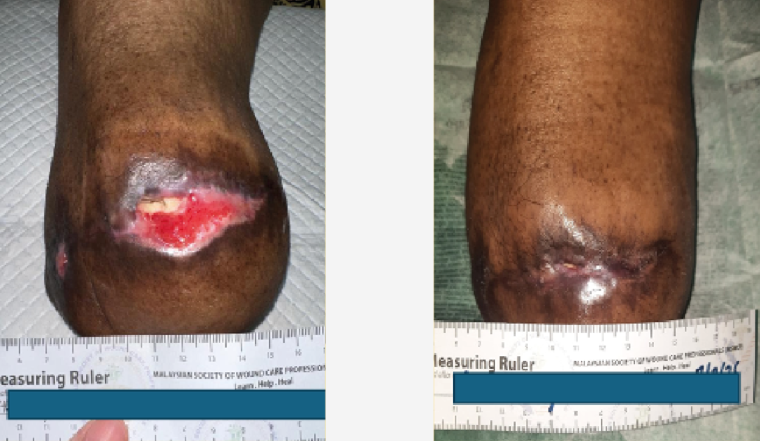
The following cases outlined in Figures 4–7 provide brief descriptive clinical examples of wound and periwound changes at baseline and endpoint of the study (8 weeks’ duration). These representative cases illustrate the wound size reduction alongside with improvement in periwound skin following application of topical MSC-CM lotion in combination with standard wound care.
Discussion
A healthy periwound skin is crucial for maintaining barrier function, modulating inflammation and serves as the initial site for epithelial migration. An observational study demonstrated that periwound maceration and barrier disruption correlate with delayed wound healing (Freitas, 2022).
Traditionally, periwound interventions are mostly based on physical protection using zinc-based products and barrier films (Perez Jaimes et al, 2025). Similarly, consensus from best practice recommendations emphasises exudate management and periwound protection with liquid barrier films as part of periwound care (LeBlanc et al, 2021).
In contrast, biologically active therapies using MSC-CM offer a regenerative approach to periwound management. Previous studies have explored MSC-based and MSC-derived therapies in wound healing, but the majority of these have focused on topical wound bed application and injectables, with limited focus on the periwound region as a therapeutic area (Sukmana et al, 2025; Sun et al, 2025).
A recent systemic review and meta-analysis highlighted that stem cell secretomes expedite diabetic wound repair by promoting angiogenesis, reducing inflammation and enhancing reepithelialisation (Suhandi et al, 2025). While this review provides strong justification for secretome-based therapies, the studies that the authors included were predominantly undertaken in animal models.
Additionally, an in vitro study using scratch assays conducted by Walter et al (2010) showed that MSC-CM facilitates migration of dermal fibroblasts and keratinocytes through paracrine signalling, supporting the findings of improved periwound skin and epithelial advancement in this case.
Further evidence exploring the key role of wound edges and surrounding epidermis in skin repair is provided by Farahat et al (2025), who demonstrated that human-induced pluripotent stem cell-derived mesenchymal stem cells significantly expedited reepithelisation in burn wound healing. Their study showed accelerated reepithelisation linked to increased keratinocyte migration from wound edges, indicating wound closure is dependent on viable keratinocytes originating from healthy adjacent skin.
The outcomes of this case series are consistent with existing evidence highlighting the regenerative potential of MSC-derived secretomes when applied to the periwound skin, rather than solely to the wound bed. These findings reinforce the understanding that wound healing extends beyond the wound bed itself and is critically influenced by the functional integrity of the surrounding periwound skin. However, this study has several limitations. As an observational case series, it lacks a control group and cannot establish causality. The sample size was small, and wounds were heterogeneous in aetiology and location. Future controlled studies incorporating validated periwound outcome measures, comparator interventions and objective functional indicators, such as TEWL measurement, are warranted to confirm these findings.
Conclusion
Adjunctive periwound application of MSC-conditioned media lotion was associated with wound area reduction and marked improvement in periwound skin. These findings support the view that periwound skin management is an essential component of overall wound care.