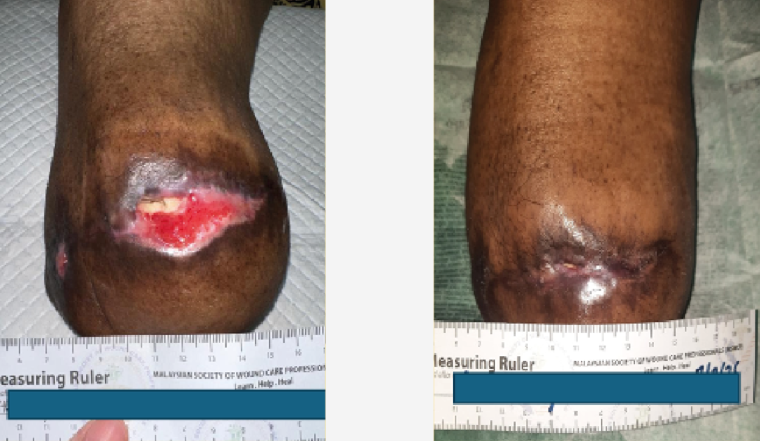
The periwound skin is defined by the International Wound Infection Institute (IWII, 2025) [Figure 1] as the skin directly adjacent to a wound. While this may appear to be a simple concept, the condition of the periwound area plays a significant and often underestimated role in wound progression and overall healing outcomes.
Promoting good periwound health can:
- Support wound healing
- Decrease the risk of wound infection
- Reduce dressing change frequency and associated cost
- Reduce pain and discomfort
- Reduce the risk of skin damage
- Improve patient comfort and quality of life (Woo et al, 2017; Whitlock et al, 2025).
When the periwound is exposed to one or more elements such as excessive moisture, wound exudate, friction, adhesive products, poor cleansing techniques or contaminants, the skin barrier can begin to break down.
This may result in maceration, inflammation, pain, epidermal stripping and an increased risk of infection or further tissue damage (World Union of Wound Healing Societies [WUWHS], 2019; Whitlock et al, 2025).
The importance of periwound health
Excess exudate is one of the most common causes of periwound skin damage. The periwound is vulnerable to moisture-associated skin damage (MASD), especially when wound drainage exceeds the fluid capacity of the dressing (Fletcher et al, 2020).
When exudate is not effectively managed, it lies against the skin, causing it to soften and become fragile. This makes the skin less tolerant to adhesives and routine care. This often creates a cycle clinicians will recognise – dressings need to be changed more frequently due to leakage, but each dressing removal may cause further trauma to already vulnerable skin.
Exudate itself can also contribute to damage. It contains inflammatory mediators, including matrix metalloproteinases (MMPs), which break down the extracellular matrix in both the wound bed and surrounding skin (Holloway et al, 2020). Proteins within wound fluid can envelop and damage corneocytes (the outermost layer of skin cells), impairing epidermal barrier function (Langøen and Bianchi, 2012). In addition, pro-inflammatory cytokines in exudate may further disrupt skin integrity and reduce barrier function (Wolcott et al, 2008).
Additional periwound damage may occur when dressings leak, exudate is not effectively managed, adhesives are repeatedly removed, or the skin is not properly cleansed and dried. Friction and shear from movement can further exacerbate skin damage.
It is also important to recognise that periwound skin may already be compromised, even if it appears intact. These subtle changes can make it more vulnerable to breakdown. Maintaining periwound integrity is therefore key, as it can directly influence whether a wound reduces in size as expected (Rippon et al, 2022).
When the periwound deteriorates, the impact is both clinical and practical. Dressings may not stay in place, leakage may increase and care becomes more demanding.
For the patient, this often means more pain, more frequent interventions and a greater overall treatment burden (LeBlanc et al, 2021).
For clinicians, the goal is not simply to absorb fluid, but to manage moisture effectively while protecting the surrounding skin. Barrier products can play an important role here, helping to shield the skin from ongoing exposure to exudate and reducing the risk of damage, particularly in patients with fragile skin or high exudate levels.
Recognising periwound damage
Regular skin assessment at every dressing change is one of the most important things a healthcare professional can do. Periwound damage often develops gradually, and early signs can be easy to miss. Identifying changes early allows for simpler, more effective and often less costly interventions.
One useful assessment framework is TIMERS (Tissue, Infection/Inflammation, Moisture balance, Edges of wound, Regeneration of tissue and Social factors; Atkin et al, 2019).
Before removing the dressing
- Take a moment to assess what you can already see and what the patient is telling you. Look for:
- Strike-through or leakage, which may indicate the dressing is no longer managing exudate effectively
- Pain around the wound, which may indicate MASD rather than pain originating from the wound bed itself.
After removing the dressing
- Assess the periwound in a structured manner. Focus on:
- Skin colour, texture, moisture level and overall condition
- Amount, colour and viscosity of exudate.
Signs of periwound damage may include:
- Macerated or fragile skin around wound edges (skin looks white, swollen, soft or waterlogged)
- Erythema or skin colour changes
- Excoriation or breakdown of the periwound area
- Sogginess or softening of the skin
- Skin drying/desiccation
- Eczema/dermatitis
- Skin denudation – loss of the epidermis
- Trauma
- Skin stripping from adhesive removal
- Infection
- Blistering
- Possible odour (LeBlanc et al, 2021; Rippon et al, 2022).
In clinical practice, any patient with a wound is at risk of periwound complications, not just those with heavily exuding wounds.
Preventing periwound complications
Prevention is always preferable to treatment. In practice, good periwound care comes down to three key steps: cleansing, protecting and selecting the right dressing – at every dressing change.
Step 1: Cleansing the periwound area
The IWII (2025) recommends that all wounds are therapeutically cleansed. This is defined as the “active removal of surface contaminants, loose debris, non-attached nonviable tissue, and/or remnants of previous dressings from the wound and periwound” (IWII, 2025).
Although evidence is insufficient to determine the ideal frequency of cleansing, the IWII (2025) supports cleansing at every dressing change. Cleansing is not simply a hygiene measure; it is a clinical intervention that directly reduces the risk of infection and periwound skin damage.
Wounds should be cleansed with an inert wound cleanser at each dressing change and before obtaining samples for microscopy, culture and sensitivity testing (IWII, 2025).
Appropriate cleansing helps remove:
- Contaminants that may migrate into the wound and increase infection risk
- Excess moisture
- Proteases within exudate that contribute to inflammation
- Adhesive residue from wound dressings that may irritate the skin and become a source of infection (IWII, 2025).
Step 2: Protecting with a barrier film
Barrier films provide an additional layer of protection for the periwound, particularly in high-exudate wounds or where adhesive trauma is a concern. Film barriers such as MedProtect Film help protect intact or at-risk skin from irritation caused by bodily fluids, exudate, adhesives and friction, while still allowing the skin to breathe.
Barrier films differ from barrier creams. This distinction is important, as barrier creams under dressings may impair adhesion, increase leakage risk and contribute to maceration. When adhesive dressings are used, barrier films are generally preferred.
Step 3: Selecting the right dressing
The periwound is susceptible to damage from multiple external sources. Dressing selection should therefore be evidence-based and focused on minimising skin trauma.
Dressings should be matched to level and type of exudate, wound size and location. If a dressing is unable to handle the volume of exudate, it can quickly become saturated, allowing fluid to leak onto the surrounding skin. On the other hand, an oversized dressing may be unnecessarily bulky and compromise adhesion.
When selecting a dressing, consider:
- Fluid-handling capacity matched to the level and type of exudate. Superabsorbent polymer (SAP) dressings (e.g. Zetuvit Plus) offer effective absorption and retention capacity in highly exuding wounds, helping to prevent leakage and strike-through. Silicone SAP dressings (such as Zetuvit Plus Silicone Border), may also be a consideration for exuding wounds (WUWHS, 2025)
- Ability to retain fluid under compression therapy, where applicable
- Prevention of leakage and strike-through
- Odour absorption where clinically indicated
- Use of atraumatic silicone adhesives (e.g. Zetuvit Plus Silicone Border) to minimise skin stripping during removal
- Correct dressing size in relation to wound dimensions
- Planned dressing change frequency according to exudate levels and dressing capacity (LeBlanc et al, 2021).
Choosing the right barrier film format
MedProtect is indicated for protecting intact or damaged skin from bodily fluids in the areas of periwound and peristomal skin protection as well as incontinence skin care.It is available in two application options to suit different clinical needs: a 28ml spray and a 1ml swab.
Additional product features include:
- Clear film, allowing ongoing skin inspection without concealing early signs of breakdown
- A film-forming product that, upon application to intact or damaged skin, forms a long-lasting waterproof barrier
- Acts as a protective interface between the skin and bodily wastes and fluids
- High moisture vapour transmission rate – moisture escapes while protection against irritants is maintained
- Dries in approximately 30 seconds, allowing dressings or adhesives to be applied promptly
- Gentle, alcohol-free and sting-free formulation suitable for sensitive or compromised skin, including in children over 12 weeks
- Compatibility with chlorhexidine gluconate (CHG) and adhesives; non-cytotoxic properties (Data on file, 2025).
MedProtect forms a long-lasting waterproof barrier that acts as a protective interface between the skin and exudate, bodily fluids and waste, while allowing the skin to breathe.
Managing periwound skin damage
Periwound skin is particularly vulnerable to maceration, excoriation and adhesive trauma. Exudate management and a consistent barrier regimen are central to protecting the healing environment.
Risk assessment and management principles
Apply the following principles at every dressing change:
- Assess the periwound skin before removing the dressing, and note any maceration, excoriation or discolouration
- Cleanse the wound and periwound gently, then pat dry
- Apply a barrier film (e.g. MedProtect Film Swab) to the periwound area before placing adhesive or dressing
- Select a wound dressing appropriate to the exudate level; consider wound size when choosing dressing size (LeBlanc et al, 2021)
- Reapply barrier film at every dressing change.
Other indications: Protecting skin at risk from body fluids
While periwound skin is the primary focus of this Made Easy, barrier films also play an important role in managing other moisture-related skin challenges commonly encountered across healthcare settings. In the conditions below, MedProtect Film can be incorporated into a structured skin protection regimen.
Incontinence-associated dermatitis (IAD)
- A form of irritant contact dermatitis caused by exposure to urine and/or faeces
- Repeated or prolonged exposure to moisture and irritants disrupts the skin barrier, increasing risk of breakdown
- Common features include diffuse erythema (or purple/maroon discolouration in darker skin tones), maceration, discomfort, burning or pain
- Severe cases may progress to skin erosion and secondary infection
- Frequently mistaken for pressure injuries in the sacral or buttock regions
- Key distinction: IAD presents as diffuse, irregular erythema with poorly defined edges and no distinct wound bed, while pressure injuries are usually localised with well-defined margins
- Accurate differentiation is essential as prevention and management differ
- Barrier films are recommended for both prevention and management of IAD (Beeckman et al, 2026).
Peristomal Moisture-Associated Dermatitis (PMAD)
- A common complication in stoma care, including around tracheostomy stomas; irritant contact dermatitis is the most frequently reported issue (Colwell et al, 2017)
- Results from exposure to stoma effluent, digestive enzymes and adhesive products
- Prevention starts with a well-fitting pouching system to minimise leakage and skin contact
- Barrier films (e.g. MedProtect Film) can be used to protect exposed peristomal skin and support ongoing skin integrity during pouch wear.
Download the PDF below to access the full Made Easy resource.